
Ears of Deep-Sea Fishes
My lab has done several studies on the ears of fishes from great depth in the oceans. As shown by images on this page, many of the features of the ears of species we have studied are rather unique, and suggest to us some kinds of “specializations” that may result in the fishes hearing particularly well, at least as compared to many species without similar adaptations. However, due to the great depth at which these species live, it has so far been impossible to actually measure their hearing. The images below, most of which were provided by my former doctoral student and colleague Dr. Xiaohong Deng, and I think her for permission to use her work. Papers from this work include:
Popper, A.N. (1980). Scanning electron microscopic studies of the sacculus and lagena in several deep‑sea fishes. Am. J. Anat. 157:115‑136
Buran, B. N., Deng, X., and Popper, A. N. (2005). Structural variation in the inner ears of four deep-sea elopomorph fishes. J. Morphol. 265:215-225.
Deng, X., Wagner, H.-J., and Popper, A. N. (2011). The inner ear and its coupling to the swim bladder in the deep-sea fish Antimora rostrata (Teleostei: Moridae). Deep Sea Research, part I, 58:27-37.
Deng, X., Wagner, H.-H., and Popper, A. N. (2013). Interspecific variations of inner ear structure in the seep-sea fish family Melamphaidae. Anatomical Record. 296, 1064-1082.
1Deng, X., Wagner, H.-J., and Popper, A. N. (2023). Comparison of the saccules and lagenae in six macrourid fishes from different deep-sea habitats). The Journal of the Acoustical Society of America, 154, 2937-2949.
All images below are courtesy of Dr. Xianong Deng from her work
on the ears of deep-sea fishes.

Inner ear of roundnoise grenadier
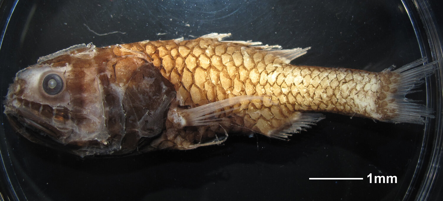
Melamphid fish - crested ridgehead

Brain and the two ears of Pain's grenadieri. Anterior to the top

Saccular otolith from a melamphaid fish. Note the elongate "rod"

SEM of a ciliary bundle from lagena of roundnose grenadier

Left image shows the saccular sensory epithelium (macula) of an Antimora. Lower image shows the hair cell orientation pattern. The arrows point in the direction of the kinocilium of the hair cells. Note that the epithelium is divided into what we refer to as hair cell orientation groups.
Drawing to the right is the utricular macula from another melaphid species. In this, and most all vertebrate utricular epithelia, the hair cells are in two major groups. There is deviation from the major orientations as the epithelia curve.


The ciliary bundles in fishes vary in length. The functional significance of different lengths is not known, but it may be related to the frequency of signals that the cells are tuned to receive. In many fishes, each region of an epithelium may have different length ciliary bundles. The variation in bundle length is seen in the utricular epithelium to the right. This is the same epithelium as the one above that has hair cell orientation patterns.
