
The Ear of Fishes
An introduction to the ear of fishes.
The ear of fishes is located in the cranial cavity just behind the eye (as seen in the drawing above from Weber, 1820). There is no external or middle ear (features of terrestrial vertebrates). Instead,, since the density of the fish body and the water is about the same, the sound field easily impinges upon the ear. In fact, the sound would travel directly through the fish and not be detected except for the presence of structures that are very different density than the rest of the body. In the ear, this is the otolith that is found in each of the end organs (see below). The other discontinuity in most bony fishes is the swim gladder, a bubble of gas (often air) int the abdominal cavity. The primary role of the swim bladder is for buoyancy control, but in many species, such as the Atlantic cod, the swim bladder is also involved in detection of the sound pressure component of the sound field.

The ear of an Atlantic cod (Gadus morhua)
The painting shows the ear with its three semicircular canals and the three otolith organs, the saccule, lagena, and utricle. The brain (in yellow) is also shown. Painting copyright by Anthony D. Hawkins and used with his kind permission. See our recent paper on hearing by Atlantic cod. Download here.
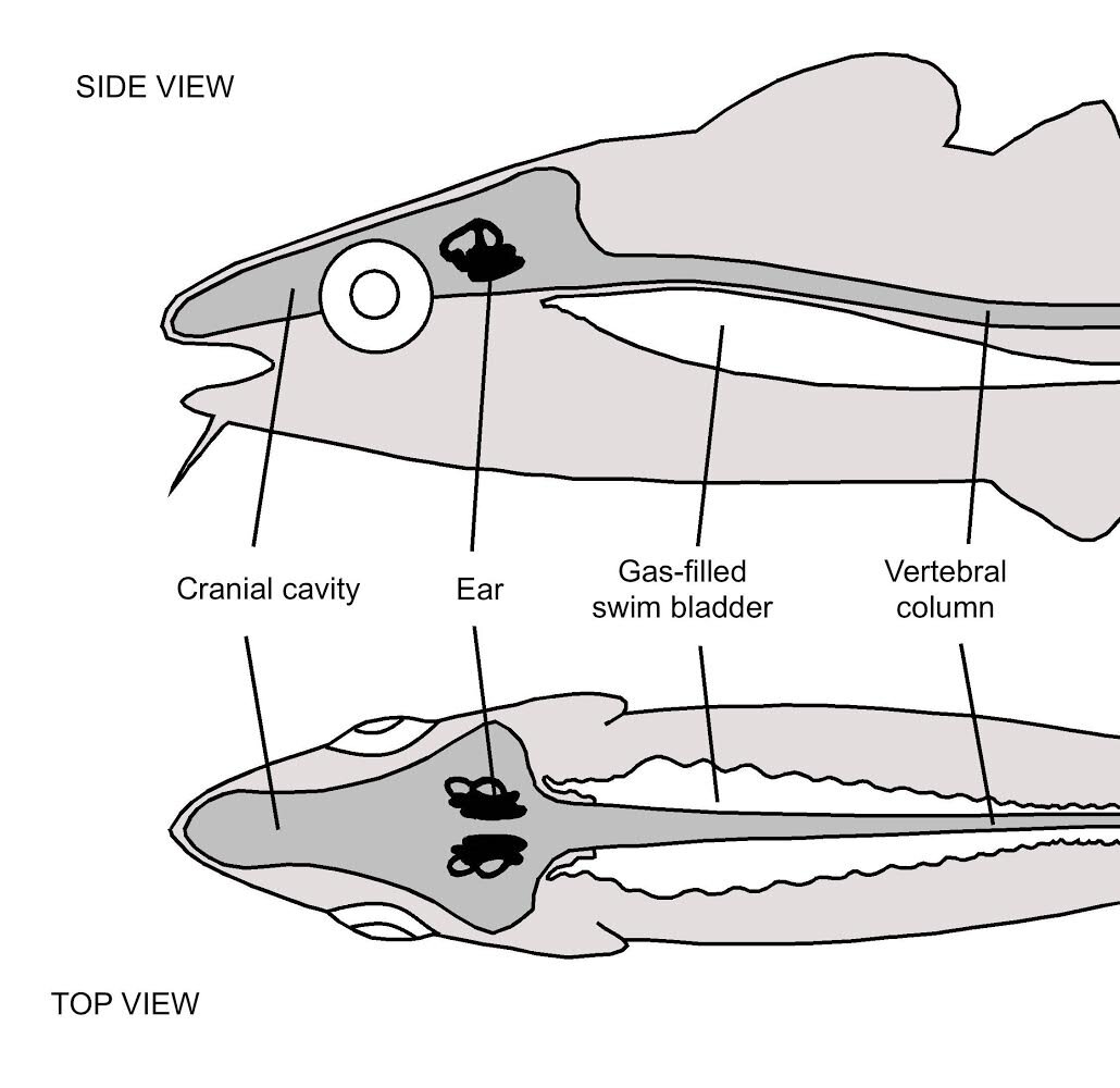
This figure shows the location of the ear in the head of the Atlantic cod. The swim bladder, a gas-filled structure in the abdominal cavity, in the cod has anterior extensions that brings it in close proximity to the ear. Figure copyright by Anthony D. Hawkins and used by permission.

Cross section of the head showing the brain and the location of the saccule. Note that the otolith lies very close to the sensory epithelium of the end organ. Figure copyright by Anthony D. Hawkins and used by permission.

Drawing of the sensory epithelium in the ear of a fish. The sensory hair cells are embedded in the sensory epithelium, and each is surrounded by supporting cells. The ciliary bundle of the sensory cells are embedded in a gelatinous otolith membrane that sits between the surface of the epithelium and the otolith. Relative motion between the epithelium and the otolith during sound stimulation causes bending of the ciliary bundles. This causes release of neurotransmitter from the cells which then stimulate the fibers of the eighth cranial nerve. That signal goes to the auditory region of the brain. Figure copyright by Anthony D. Hawkins and used by permission.

Scanning electron micrograph (SEM) of ciliary bundles on sensory hair cells of a fish saccular epithelium (macula). Note that the ciliary bundles are “polarized" morphologically, so that the longest cilium, called the kinocilium, is at one end of the bundle. The other cilia, called strereocilia, are often graded in size..

The response of sensory hair cells, based on the work of my late friend, Professor Ake Flock. Sensory hair cells are innervated by fibers of the eighth nerve. When the ciliary bundles are bent in different direction they release different amounts of neurotransmitter to stimulate the nerve fibers. When the bundles are bent towards the kinocilium they give out maximum response, and they have minimal responses in the opposite direction. As can be seen in the drawing on the right, each direction results in a different level of stimulation of the nerve. This directional response property is the basis for fishes doing sound source localization. Figure copyright by Anthony D. Hawkins and used by permission.

Otoliths from the saccule of various fish species. Otolith shape is species-specific, and fisheries biologists and taxonomists use the otoliths to identify species, even in the fossil record. The groove in each otolith is the location of the saccular sensory epithelium. In each case, anterior is to the left and dorsal to the top.

Scanning electron micrograph showing the saccular epithelium from a fish. Note the otolithic membrane at the top of the image.

Schematic of the sensory hair cells from the saccule in a fish. This drawing shows details of the two types of cells found in fishes..

In fishes called the Otophysans, a large fresh water group that includes catfish, goldfish, zebrafish, and almost 5,000 other species, the anterior end of the swim bladder connects to a series of bones, the Weberian ossicles. When the wall of the swim bladder moves in response to sound, the ossicles move (analogously to the mammalian middle ear bones) and stimulates the inner ear. This results in these fishes hearing a wider range of frequencies (bandwidth) and lower sound levels than most fishes without the ossicles.